A groundbreaking study by Nankai University scientists reveals a novel method for synthesizing quantum dots within the nuclei of live cells. This technique, leveraging the cell’s natural processes with glutathione, paves the way for advanced applications in synthetic biology, including the production of nanodrugs and nanorobots, by enabling precise inorganic material synthesis at the subcellular level.
A recent study published in the journal National Science Review demonstrates the synthesis of quantum dots (QDs) in the nucleus of live cells. The research was conducted by Dr. Hu Yusi, Associate Professor Wang Zhi-Gang, and Professor Pang Dai-Wen from Nankai University.
During the study of QDs synthesis in mammalian cells, it was found that the treatment with glutathione (GSH) enhanced the cell’s reducing capacity. The generated QDs were not uniformly distributed within the cell but concentrated in a specific area. Through a series of experiments, it was confirmed that this area is indeed the cell nucleus (as shown in the figure). Dr. Hu said, “This is truly amazing, almost unbelievable.”
Understanding the Molecular Mechanisms
Dr. Hu and his mentor Professor Pang attempted to elucidate the molecular mechanism of quantum dot synthesis in the cell nucleus. It was found that GSH plays a significant role. There is a GSH transport protein, Bcl-2, on the nucleus, which transports GSH into the nucleus in large quantities, enhancing the reducing ability within the nucleus, promoting the generation of Se precursors. At the same time, GSH can also expose thiol groups on proteins, creating conditions for the generation of Cd precursors. The combination of these factors ultimately enables the abundant synthesis of quantum dots in the cell nucleus.
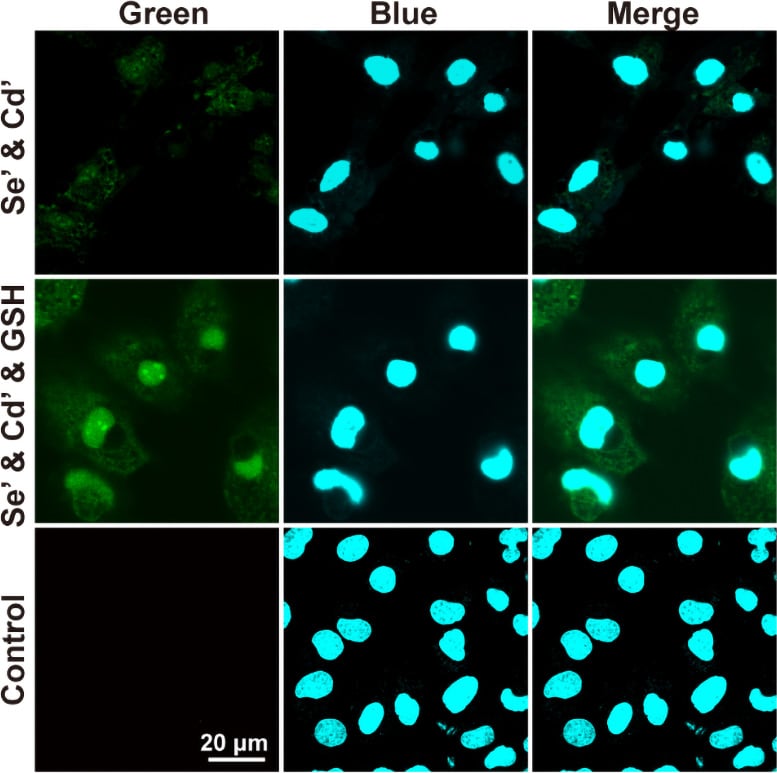
From left to right, the fluorescence images of the QDs, the fluorescence images of the nucleus staining dye and the merge of the two. This figure shows that with the treatment of GSH, the fluorescent QDs were grown in the nucleus of live cells. Se’ stands for Na2SeO3; Cd’ stands for CdCl2. Credit: Science China Press
Professor Pang stated, “This is an exciting result; this work achieves the precise synthesis of QDs in live cells at the subcellular level.” He continued, “Research in the field of synthetic biology mostly focuses on live cell synthesis of organic molecules through reverse genetics. Rarely do we see the live cell synthesis of inorganic functional materials. Our study doesn’t involve complex genetic modifications; it achieves the target synthesis of inorganic fluorescent nanomaterials in cellular organelles simply by regulating the content and distribution of GSH within the cell. This addresses the deficiency in synthetic biology for the synthesis of inorganic materials.”
While the synthesis of organic materials in cells remains predominant in the field of biosynthesis, this research undoubtedly paves the way for the synthesis of inorganic materials in synthetic biology. Professor Pang expressed, “Each of our advancements is a new starting point. We firmly believe that in the near future, we can use cell synthesis to produce nanodrugs, or even nanorobots in specified organelles. Moreover, we can transform cells into super cells, enabling them to do unimaginable things.”
Reference: “In-situ synthesis of quantum dots in the nucleus of live cells” by Yusi Hu, Zhi-Gang Wang, Haohao Fu, Chuanzheng Zhou, Wensheng Cai, Xueguang Shao, Shu-Lin Liu and Dai-Wen Pang, 12 January 2024, National Science Review.
DOI: 10.1093/nsr/nwae021

Dr. Thomas Hughes is a UK-based scientist and science communicator who makes complex topics accessible to readers. His articles explore breakthroughs in various scientific disciplines, from space exploration to cutting-edge research.