Summary: A new study reveals a vital brain-fat tissue feedback loop that plays a pivotal role in aging. The research identifies specific neurons in the hypothalamus that, when activated, signal the body’s fat tissue to release energy, facilitating physical activity and brain function. As this feedback loop deteriorates with age, health problems associated with aging become more prevalent.
Mice with a constantly active feedback loop displayed delayed aging, increased physical activity, and longer lifespans. This groundbreaking research offers potential insights for future interventions in aging and longevity.
Key Facts:
- The study unveils a critical brain-fat tissue feedback loop that influences aging and health.
- Activation of specific neurons in the hypothalamus triggers the release of fatty acids and enzymes that fuel the body and brain.
- Mice with a sustained feedback loop lived longer, were more physically active, and displayed signs of delayed aging.
Source: WUSTL
In recent years, research has begun to reveal that the lines of communication between the body’s organs are key regulators of aging. When these lines are open, the body’s organs and systems work well together. But with age, communication lines deteriorate, and organs don’t get the molecular and electrical messages they need to function properly.
A new study from Washington University School of Medicine in St. Louis identifies, in mice, a critical communication pathway connecting the brain and the body’s fat tissue in a feedback loop that appears central to energy production throughout the body. The research suggests that the gradual deterioration of this feedback loop contributes to the increasing health problems that are typical of natural aging.
The study — published Jan. 8 in the journal Cell Metabolism — has implications for developing future interventions that could maintain the feedback loop longer and slow the effects of advancing age.
The researchers identified a specific set of neurons in the brain’s hypothalamus that, when active, sends signals to the body’s fat tissue to release energy. Using genetic and molecular methods, the researchers studied mice that were programmed to have this communication pathway constantly open after they reached a certain age.
The scientists found that these mice were more physically active, showed signs of delayed aging, and lived longer than mice in which this same communication pathway gradually slowed down as part of normal aging.
“We demonstrated a way to delay aging and extend healthy life spans in mice by manipulating an important part of the brain,” said senior author Shin-ichiro Imai, MD, PhD, the Theodore and Bertha Bryan Distinguished Professor in Environmental Medicine and a professor in the Department of Developmental Biology at Washington University.
“Showing this effect in a mammal is an important contribution to the field; past work demonstrating an extension of life span in this way has been conducted in less complex organisms, such as worms and fruit flies.”
These specific neurons, in a part of the brain called the dorsomedial hypothalamus, produce an important protein — Ppp1r17. When this protein is present in the nucleus, the neurons are active and stimulate the sympathetic nervous system, which governs the body’s fight or flight response.
The fight or flight response is well known for having broad effects throughout the body, including causing increased heart rate and slowed digestion. As part of this response, the researchers found that the neurons in the hypothalamus set off a chain of events that triggers neurons that govern white adipose tissue — a type of fat tissue — stored under the skin and in the abdominal area.
The activated fat tissue releases fatty acids into the bloodstream that can be used to fuel physical activity. The activated fat tissue also releases another important protein — an enzyme called eNAMPT — which returns to the hypothalamus and allows the brain to produce fuel for its functions.
This feedback loop is critical for fueling the body and the brain, but it slows down over time. With age, the researchers found that the protein Ppp1r17 tends to leave the nucleus of the neurons, and when that happens, the neurons in the hypothalamus send weaker signals.
With less use, the nervous system wiring throughout the white adipose tissue gradually retracts, and what was once a dense network of interconnecting nerves becomes sparse. The fat tissues no longer receive as many signals to release fatty acids and eNAMPT, which leads to fat accumulation, weight gain and less energy to fuel the brain and other tissues.
The researchers, including first author Kyohei Tokizane, PhD, a staff scientist and a former postdoctoral researcher in Imai’s lab, found that when they used genetic methods in old mice to keep Ppp1r17 in the nucleus of the neurons in the hypothalamus, the mice were more physically active — with increased wheel-running — and lived longer than control mice. They also used a technique to directly activate these specific neurons in the hypothalamus of old mice, and they observed similar anti-aging effects.
On average, the high end of the life span of a typical laboratory mouse is about 900 to 1,000 days, or about 2.5 years. In this study, all of the control mice that had aged normally died by 1,000 days of age. Those that underwent interventions to maintain the brain-fat tissue feedback loop lived 60 to 70 days longer than control mice.
That translates into an increase in life span of about 7%. In people, a 7% increase in a 75-year life span translates to about five more years. The mice receiving the interventions also were more active and looked younger — with thicker and shinier coats — at later ages, suggesting more time with better health as well.
Imai and his team are continuing to investigate ways to maintain the feedback loop between the hypothalamus and the fat tissue. One route they are studying involves supplementing mice with eNAMPT, the enzyme produced by the fat tissue that returns to the brain and fuels the hypothalamus, among other tissues.
When released by the fat tissue into the bloodstream, the enzyme is packaged inside compartments called extracellular vesicles, which can be collected and isolated from blood.
“We can envision a possible anti-aging therapy that involves delivering eNAMPT in various ways,” Imai said.
“We already have shown that administering eNAMPT in extracellular vesicles increases cellular energy levels in the hypothalamus and extends life span in mice. We look forward to continuing our work investigating ways to maintain this central feedback loop between the brain and the body’s fat tissues in ways that we hope will extend health and life span.”
Funding: This work was supported by the National Institute on Aging of the National Institutes of Health (NIH), grant numbers AG037457 and AG047902; the American Federation for Aging Research; the Tanaka Fund at Washington University School of Medicine; a Glenn Foundation for Medical Research Postdoctoral Fellowship; and a Tanaka Scholarship. The content is solely the responsibility of the authors and does not necessarily represent the official views of the NIH.
About this aging and neuroscience research news
Author: Diane Williams
Source: WUSTL
Contact: Diane Williams – WUSTL
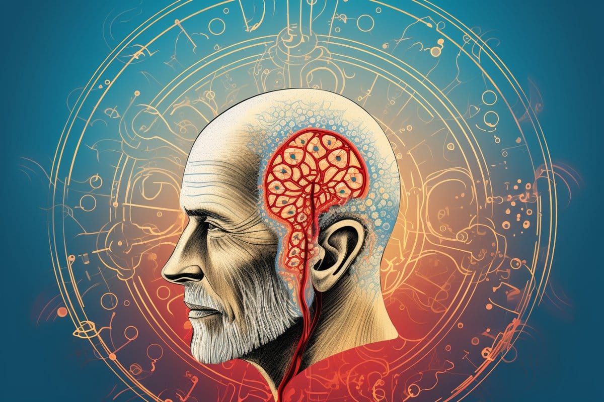
Image: The image is credited to Neuroscience News
Original Research: Open access.
“DMHPpp1r17 neurons regulate aging and lifespan in mice through hypothalamic-adipose inter-tissue communication” by Shin-ichiro Imai et al. Cell Metabolism
Abstract
DMHPpp1r17 neurons regulate aging and lifespan in mice through hypothalamic-adipose inter-tissue communication
Recent studies have shown that the hypothalamus functions as a control center of aging in mammals that counteracts age-associated physiological decline through inter-tissue communications.
We have identified a key neuronal subpopulation in the dorsomedial hypothalamus (DMH), marked by Ppp1r17 expression (DMHPpp1r17 neurons), that regulates aging and longevity in mice. DMHPpp1r17 neurons regulate physical activity and WAT function, including the secretion of extracellular nicotinamide phosphoribosyltransferase (eNAMPT), through sympathetic nervous stimulation.
Within DMHPpp1r17 neurons, the phosphorylation and subsequent nuclear-cytoplasmic translocation of Ppp1r17, regulated by cGMP-dependent protein kinase G (PKG; Prkg1), affect gene expression regulating synaptic function, causing synaptic transmission dysfunction and impaired WAT function.
Both DMH-specific Prkg1 knockdown, which suppresses age-associated Ppp1r17 translocation, and the chemogenetic activation of DMHPpp1r17 neurons significantly ameliorate age-associated dysfunction in WAT, increase physical activity, and extend lifespan.
Thus, these findings clearly demonstrate the importance of the inter-tissue communication between the hypothalamus and WAT in mammalian aging and longevity control.

Sarah Carter is a health and wellness expert residing in the UK. With a background in healthcare, she offers evidence-based advice on fitness, nutrition, and mental well-being, promoting healthier living for readers.


